
Digital X-rays in Cordova, TN
Digital X-rays let Dr. Dalal see what a visual exam cannot, with a fraction of the radiation of older film. We use them to catch small problems early and to explain, in plain language, what is happening in your mouth.
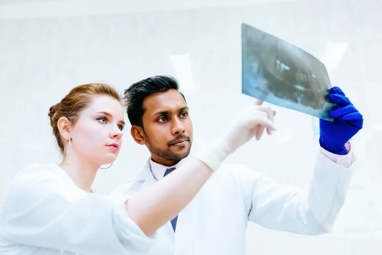
What to Know About Digital X-rays in Cordova TN
If you have ever sat in a dental chair and wondered what the doctor was actually looking at on the screen, this page is for you. Digital X-rays in Cordova TN are part of almost every exam at Cordova Family Dental, and we use them the way a good mechanic uses a flashlight: to see the parts of the tooth and bone that we cannot see with the eye alone. Then we turn the screen toward you and show you what we found.
Dr. Dalal trained at the University of Tennessee College of Dentistry and has practiced in Memphis, Kingsport, and Union City. Across all of those communities, the patients who felt most comfortable with their care were the ones who understood the images on the monitor. That is why we walk through every X-ray with you before we recommend anything.
Our office uses sensors and software that produce a clear image in seconds, so there is no waiting on chemicals or film. The image lives in your chart, and we can pull it up at your next visit to compare. That comparison is often how we catch a small cavity or a hairline fracture before it turns into a bigger problem.
How Digital X-rays Differ From Older Film X-rays
The biggest practical difference is radiation. Digital sensors need far less exposure than traditional film to produce a usable image, which matters when we are taking routine bitewings on a child or doing a full series for a new adult patient. A typical set of digital bitewings exposes you to a small fraction of what you absorb on a cross-country flight.
The second difference is speed and clarity. The image appears on the monitor right next to you in a few seconds, and we can zoom in, adjust contrast, and measure. If Dr. Dalal sees a shadow between two molars, she can enlarge that exact area and show you what she is seeing instead of describing it in the abstract.
The third piece is the software layer. We read our digital X-rays with OverJet AI assistance, which highlights areas the algorithm flags for a closer look. Dr. Dalal still makes every diagnosis herself. The AI is a second set of eyes, not a replacement, and it is part of how we keep things from slipping through the cracks.
When You Need X-rays and How Often We Recommend Them
For most adults with stable dental health, bitewing X-rays once a year and a full mouth series every three to five years is a reasonable rhythm. If you have a history of cavities, gum disease, or recent restorative work, we may take images more often so we can keep an eye on specific teeth. We will tell you why before we take anything.
For kids, we usually start with a small number of images once the back teeth are touching, since that is when cavities can hide between them. We never take X-rays on a child just because they are due. There has to be a reason, and we will explain that reason to the parent in the room.
There are also focused situations where a single image answers a specific question: a tooth that has been bothering you, a suspected crack, a deep filling that may be close to the nerve, or planning for a crown or an extraction. For implant planning and complex cases, we step up to CBCT 3D imaging, which gives us a three-dimensional view of the bone.
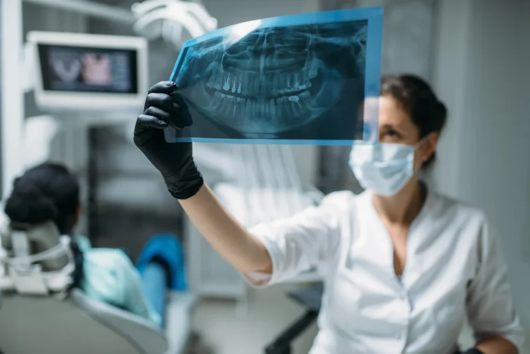
What Happens During a Digital X-ray Appointment
When you sit down for X-rays at our Cordova office, the assistant will drape a lead apron with a thyroid collar over you. The sensor is a small, flat piece that sits inside your mouth while we position the arm of the X-ray machine outside your cheek. Each image takes a fraction of a second.
For a routine set of bitewings, plan on about five minutes from start to finish. A full mouth series takes a little longer because we are capturing every tooth from several angles. A panoramic image, which sweeps around the outside of your head, is even faster and does not require sensors inside the mouth.
Once the images are on the screen, Dr. Dalal will sit down with you and walk through them. If everything looks healthy, she will tell you that plainly. If she sees something, she will point to it, explain what it means, and lay out the options, whether that is watching and waiting, a filling, or a referral. The goal is that you leave understanding the why, not just the what.
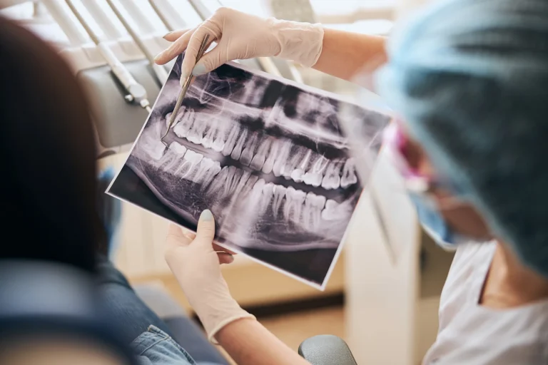
Why Digital Imaging Changes What We Can Catch Early
Most of the conditions we treat are easier, less expensive, and more comfortable to address when they are small. A cavity caught between two molars on a bitewing is a routine filling. The same cavity, missed for two years, can become a crown or a root canal. Digital imaging is how we keep small problems small.
It also gives you a record over time. When you come in next year, we can put this year’s images next to last year’s and see whether a watched area has changed. That kind of side-by-side comparison is hard to do well with film, and it is one of the quietest but most useful parts of how we practice.
If you want to see the rest of what we use chairside, our page on dental technology in Cordova TN covers the intraoral scanner, the intraoral camera, and our CBCT. Together they let us show you your mouth on a screen instead of asking you to take our word for it.
Ready for an Exam That Includes Digital X-rays?
Whether you are due for a checkup or you have a specific tooth on your mind, call Cordova Family Dental and we will get you on the schedule. New patients from Cordova, Bartlett, Lakeland, Germantown, and east Memphis are welcome.